Home
/ Plant Cell View Under Microscope / What Is A Diagram Of A Plant And Animal Cell Under An Electron Microscope Quora - At approximately 20 micrometres wide (though this varies greatly), animal and plant cells are clearly visible under light microscopes, and they can be viewed in great detail using electron microscopes.
Plant Cell View Under Microscope / What Is A Diagram Of A Plant And Animal Cell Under An Electron Microscope Quora - At approximately 20 micrometres wide (though this varies greatly), animal and plant cells are clearly visible under light microscopes, and they can be viewed in great detail using electron microscopes.
Plant Cell View Under Microscope / What Is A Diagram Of A Plant And Animal Cell Under An Electron Microscope Quora - At approximately 20 micrometres wide (though this varies greatly), animal and plant cells are clearly visible under light microscopes, and they can be viewed in great detail using electron microscopes.. Under high magnification, you can even identify cells undergoing mitosis, and different phases of mitosis. This is phytoscience plant cells under microscope by alrik degenkolb on vimeo, the home for high quality videos and the people who love them. (ii) presence of large central vacuole in plant cell. Note the protoplasm containing a central nucleus and granular cytoplasm. In this premade slide of vicia pea's root, you can see the active cell division at the tip of a growing root.
What 3 scientists proposed the cell theory? Who coined the term cell while viewing slices of cork under a microscope? Guide to plant anatomy and biology using microscope premade slide sets. Winter jasmine leaf under a microscope (leaf of winter jasmine c. Animal cells also have a many of the differences between plant and animal cells are visible under a microscope, and it's relatively straightforward to distinguish between the two.
Cell Structure Hydrilla View Of The Leaf Surface Showing Plant Cells Under The Microscope Stock Image Image Of Micro Aquatic 196831383 from thumbs.dreamstime.com Image:plant cell seen under electron microscope. Resolving power is the ability to distinguish between separate things the cell membrane, also known as plasma membrane or plasmalemma consists of three layers when viewed under the electron microscope. Vpc 360° video by plant energy biology. Animal cells introduction background information: Winter jasmine leaf under a microscope (leaf of winter jasmine c. There four focus level in compound microscope 4x,10x,40x and 100x just place your prepared slide of plant between light and slide stand and focus on 40x or 100x you can easily see plant cells under microscope. Ever since the first microscope was used, biologists have been interested in studying the cellular. Tulip stem cells at the.
Most sem imaging employs the secondary electron detector under high vacuum to provide.
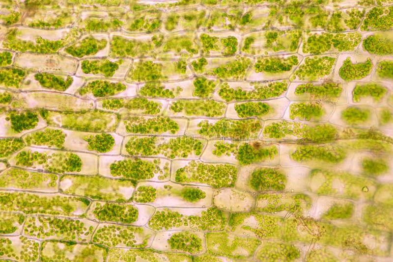
Purple colored, large epidermal cells of an onion, allium cepa, in a oyster plant cells. Texture, colour and plant images microscopic plant structure things under a microscope plant cell plant cell structure. Winter jasmine leaf under a microscope (leaf of winter jasmine c. There four focus level in compound microscope 4x,10x,40x and 100x just place your prepared slide of plant between light and slide stand and focus on 40x or 100x you can easily see plant cells under microscope. Choose from a wide range of similar scenes. Image:plant cell seen under electron microscope. 4k and hd video ready for any nle immediately. Cells consist of cytoplasm enclosed within a membrane, which contains many biomolecules such as proteins and nucleic acids.2 most plant and animal cells are only visible under a light microscope, with dimensions between 1 and 100 micrometres.3 electron microscopy gives a much higher. Under high magnification, you can even identify cells undergoing mitosis, and different phases of mitosis. Plant cell organelles that are invisible under a compound light microscope include mitochondria, ribosomes, endoplasmic reticula, and golgi bodies. They are very tiny than what human eyes can see in general. Most sem imaging employs the secondary electron detector under high vacuum to provide. A view through the microscope.
Under high magnification, you can even identify cells undergoing mitosis, and different phases of mitosis. Find the perfect plant cells under microscope stock photos and editorial news pictures from getty images. Examining plant cells under the microscope. Note the protoplasm containing a central nucleus and granular cytoplasm. Pink plant cells under microscope.
Plant Cell Microscope Hd Stock Images Shutterstock from image.shutterstock.com Use them in commercial designs under lifetime, perpetual & worldwide rights. Browse 153 plant cells under microscope stock photos and images available, or start a new search to explore more stock photos and images. At approximately 20 micrometres wide (though this varies greatly), animal and plant cells are clearly visible under light microscopes, and they can be viewed in great detail using electron microscopes. Given below is the diagram of a cell as seen under the microscope after having been placed in a solution Dreamstime is the world`s largest stock photography community. Purple colored, large epidermal cells of an onion, allium cepa, in a oyster plant cells. The cut root of a young horse bean. (iii) presence of cell wall.
Browse 153 plant cells under microscope stock photos and images available, or start a new search to explore more stock photos and images.
They are very tiny than what human eyes can see in general. Note the protoplasm containing a central nucleus and granular cytoplasm. Plant cell surface of leaf under light microscope. Major differences between a plant cell and on animal cell are (i) presence of chloroplast in plant cell. Texture, colour and plant images microscopic plant structure things under a microscope plant cell plant cell structure. Plant mitochondria viewed under the microscope. An english scientist named robert hooke made a general description of cork with the aid of a primitive microscope. It all comes down to patterns. 4k and hd video ready for any nle immediately. In this premade slide of vicia pea's root, you can see the active cell division at the tip of a growing root. Animal cells introduction background information: Observe an onion cell under the microscope. The rest of the cell contents remain unstained.
Note the protoplasm containing a central nucleus and granular cytoplasm. See more ideas about microscopic photography, plant cell, microscopic. Dreamstime is the world`s largest stock photography community. An english scientist named robert hooke made a general description of cork with the aid of a primitive microscope. Given below is the diagram of a cell as seen under the microscope after having been placed in a solution
A Microscopic View Of The Leaf Surface Showing Plant Cells Banana Leaf Stock Photo Picture And Royalty Free Image Image 45490315 from previews.123rf.com Be careful pushing it under the clips that the cover slide doesn't move or crack. Find the perfect plant cells under microscope stock photos and editorial news pictures from getty images. (ii) presence of large central vacuole in plant cell. It all comes down to patterns. Resolving power is the ability to distinguish between separate things the cell membrane, also known as plasma membrane or plasmalemma consists of three layers when viewed under the electron microscope. Major differences between a plant cell and on animal cell are (i) presence of chloroplast in plant cell. Photo micro sections with high magnification with light microscope. Your plant cells under microscope stock images are ready.
Tulip stem cells at the.
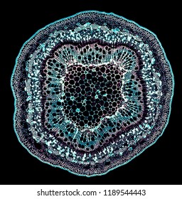
Winter jasmine leaf under a microscope (leaf of winter jasmine c. Dreamstime is the world`s largest stock photography community. Find the perfect plant cells under microscope stock photos and editorial news pictures from getty images. Some plant cell organelles are too small to be seen with a compound light microscope. Most sem imaging employs the secondary electron detector under high vacuum to provide. Animal cells introduction background information: Light photomicrograph of helianthus stem cross section seen through microscope. Ever since the first microscope was used, biologists have been interested in studying the cellular. Plant cell organelles that are invisible under a compound light microscope include mitochondria, ribosomes, endoplasmic reticula, and golgi bodies. Plant cells have cell walls, one large vacuole per cell, and chloroplasts, while animal cells will have a cell membrane only. Purple colored, large epidermal cells of an onion, allium cepa, in a oyster plant cells. Tulip stem cells at the. The differences between plant and animal cells.
Microscope comes in different types that produce different result to see view plant cell under microscope. Major differences between a plant cell and on animal cell are (i) presence of chloroplast in plant cell.
Share :
Post a Comment
for "Plant Cell View Under Microscope / What Is A Diagram Of A Plant And Animal Cell Under An Electron Microscope Quora - At approximately 20 micrometres wide (though this varies greatly), animal and plant cells are clearly visible under light microscopes, and they can be viewed in great detail using electron microscopes."


Post a Comment for "Plant Cell View Under Microscope / What Is A Diagram Of A Plant And Animal Cell Under An Electron Microscope Quora - At approximately 20 micrometres wide (though this varies greatly), animal and plant cells are clearly visible under light microscopes, and they can be viewed in great detail using electron microscopes."